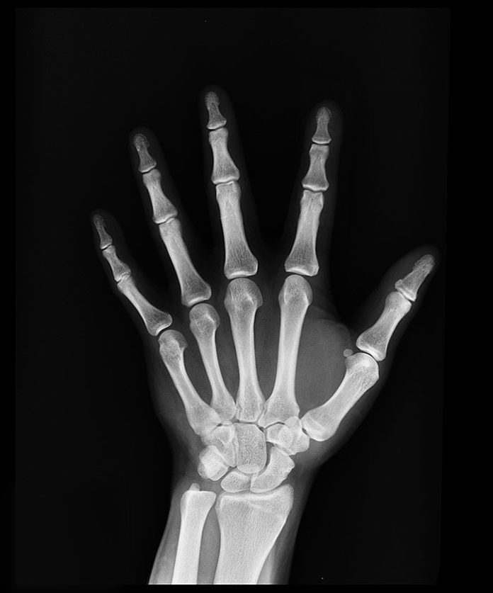
Although bones are rigid, they can bend or “give” a little if required due to outside force. However, there’s obviously a limit and a force that’s too strong for a bone may lead to a fracture.
How severe a fracture is generally depends on the strength and angle of the blow that caused it. If the breaking point of the bone has only been exceeded very slightly, it may simply crack rather than break right the way through. If the force is very violent when sustained however, such as via a gunshot wound or a road traffic accident, the bone may shatter altogether.
Once a break has occurred, some bone fragments may stick out through the skin, or the wound may go down deeply to the broken bone. This is an “open” fracture. Open fractures are particularly serious because broken skin can allow for infection to take hold, both in the bone and the wound.
Common types of fractures include:
- Stable fractures. This is where the ends of the broken bone line up and are only slightly out of place.
- Open, compound fracture. This is where the bone has pierced through the skin.
- Oblique fracture. This means the break is at an angle.
- Transverse fracture. This type of break has a fracture line which is horizontal.
- Comminuted fracture. This is where the bone has shattered into three or more pieces.
Causes
The most common causes of fractures are trauma (such as accidents or falls), overuse (stress fractures for example), or Osteoporosis (particularly in older women).
Symptoms
Many fractures are very painful and will impair movement. Other common symptoms include tenderness and swelling around the injury, bruising, deformity or a part of the bone puncturing through the skin. These symptoms are very unpleasant and can leave permanent damage so it’s vital that the type of fracture a patient has is diagnosed quickly and effectively so treatment can begin.
Learn accurate X-ray interpretation skills with our recognised CDP course
Our X-ray interpretation of minor injuries workshop (including Red Dot) examines the different types of breaks in detail, how they present on X-ray and how to interpret them. Patient assessment, radiographic referral, interpretation and treatment for minor injuries are all included.
Some experience with x-ray interpretation is required, although all equipment, course materials and refreshments are provided.
Courses last for one day and take place at Hamilton House, London, on 22nd November 2019 and 21st May 2020. The course is worth 7 hours of CPD. Places are limited so make sure you secure your space today.


